CLINICAL CANCER OF THE INTEGUMENTARY SYSTEM

Cancer of the skin is a disease with high prevalence worldwide. Melanoma in advanced stages comes with significant morbidity and risk of mortality. However, the less dangerous non-melanoma skin cancers provide clinical areas of improvement. In a variety of clinical trials, optoacoustic imaging has shown the potential to improve cancer staging (e.g. by non-invasive assessment of Breslow depth and sentinel lymph node involvement in melanoma) and surgery planning (e.g. for Mohs surgery in basal cell carcinoma).

Volumetric MSOT image of a basal cell carcinoma, visualizing spectrally unmixed melanin (yellow), oxygenated hemoglobin (red), and deoxygenated hemoglobin (blue).

3D imaging of non-melanoma skin cancer

A handheld hemispherical detector can be used to visualize melanotic or non-melanotic skin cancers, as even non-melanotic skin lesions can be sufficiently pigmented to allow non-contrast-enhanced detection via MSOT. Knowing the dimensions of a lesion prior to surgery using MSOT could potentially shorten the duration of Mohs’ micrographic surgery.

Chuah et al. J Invest Dermatol. 2019

Non-invasive assessment of tumor dimensions

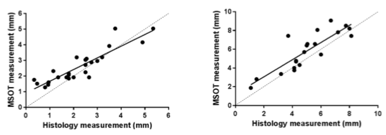
Measurements of depth (left) and length (right) acquired by MSOT are consistent with histological measurements performed after biopsy. MSOT could potentially provide a non-invasive means of determining key morphological features of non-melanotic skin cancers.

Chuah et al. J Invest Dermatol. 2019

Correlation of volumetric MSOT measurements of non-melanoma skin cancer with histology, showing depth (left) and length correlations (right).