Peripheral arterial disease (PAD) results from a buildup of atherosclerotic plaques in the blood vessels of the legs, resulting in narrowing of the arteries and decreased blood flow in the muscles and surrounding tissues. Current diagnostic methods can visualize anatomical features (like blood vessels or stenosis), quantify blood flow or measure microcirculation of the skin. However, these techniques cannot assess microcirculation in the muscle, a critical parameter for disease progression. Optoacoustic imaging has shown the potential to quantify the perfusion and oxidative capacity in muscle tissue as a biomarker of ischemia.
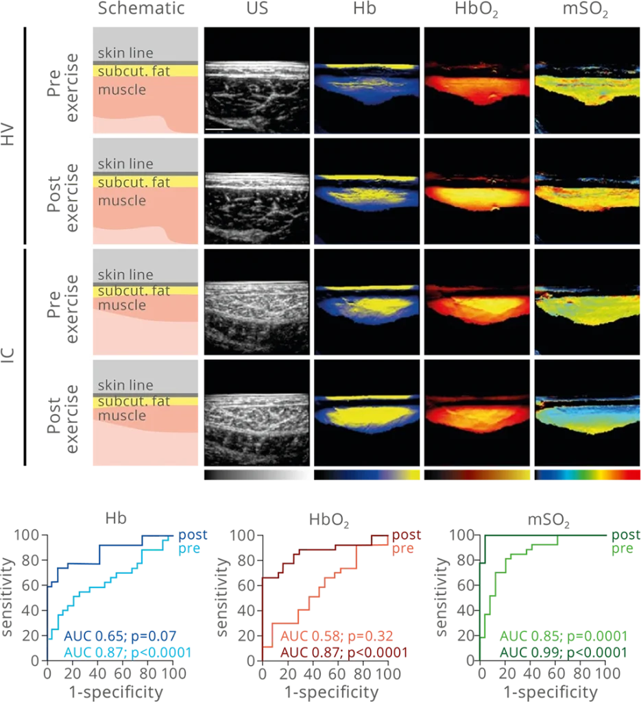
Spectrally unmixed MSOT signals show oxygenated hemoglobin (yellow to red), deoxygenated hemoglobin (yellow to blue) and MSOT-derived oxygen saturation (blue to red). ROC of MSOT signals are quantified before and after provocation. Ultrasound signal shown in grayscale.
Oxidative capacity as a biomarker of disease severity
Intermittent claudication (IC) patients and healthy volunteers (HV) were enrolled in a prospective study to evaluate the diagnostic accuracy by MSOT. MSOT-derived oxygen saturation after exercise shows a sensitivity and a specificity of 100% and 95.8%, respectively. These results show the potential of MSOT-derived saturation as a diagnostic biomarker to distinguish between HV and IC patients.
Caranovic M et al. medRxiv. 2023
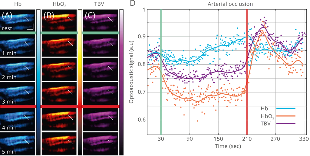
Following arterial occlusion in healthy volunteers
Healthy volunteers (HV) were enrolled in a case study to evaluate the potential of MSOT to detect and follow muscle hemodynamics. Post-occlusion hyperemic response of oxygenated hemoglobin was observed. These results show the potential of MSOT as a tool to measure vascular changes in the affected limb, which with further validation could potentially identify high-risk PAD patients by assessing microvascular dysfunction.
Karlas A et al. J Biophotonics. 2020
Evaluation of muscle hemodynamics using MSOT before, during and after arterial occlusion. Spectrally unmixed oxygenated hemoglobin (red to white), deoxygenated hemoglobin (blue to white), and total hemoglobin (purple to white) is quantified over time.
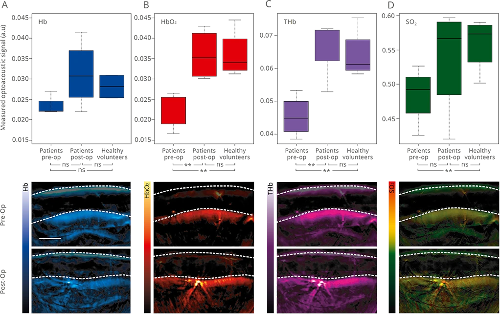
Representative MSOT images from PAD patients (IC) and healthy volunteers (HV), visualizing oxygenated hemoglobin (red to yellow), deoxygenated hemoglobin (blue to white), total hemoglobin (purple to white) and MSOT-derivedoxygensaturation (blacktored). MSOT signals were quantified before and after endovascular revascularization and compared to HV.
Impact of revascularization in PAD patients
Intermittent claudication (IC) patients and healthy volunteers (HV) were enrolled in a case study to evaluate the difference in perfusion and oxidative capacity between the two groups. The impact of interventional therapy on muscle hemodynamics was studied. The results show the potential of MSOT to visualize and quantify the effect of therapy using functional parameters of muscle perfusion and oxidative capacity.