Optoacoustic imaging has the unique ability to directly visualize and quantify the molecular composition of tissue, based on the ability of various biomarkers to absorb light. This is made possible by the photoacoustic effect – the conversion of light energy into acoustic signals.
Non-invasive optical imaging, at depth.
With a non-invasive procedure, real-time high-resolution image display, and reaching depths of up to 3 cm in tissue, optoacoustic imaging has demonstrated unique clinical and scientific value in numerous clinical and preclinical applications.
Real-time imaging chain
After acquisition, the raw data is reconstructed to form images. Spectral analysis then separates the contribution of different chromophores – both intrinsic (e.g., hemoglobin, collagen, melanin, water, lipids) and extrinsic (e.g., indocyanine green (ICG), nanoparticles). Finally, anatomical features are enhanced and visualized.
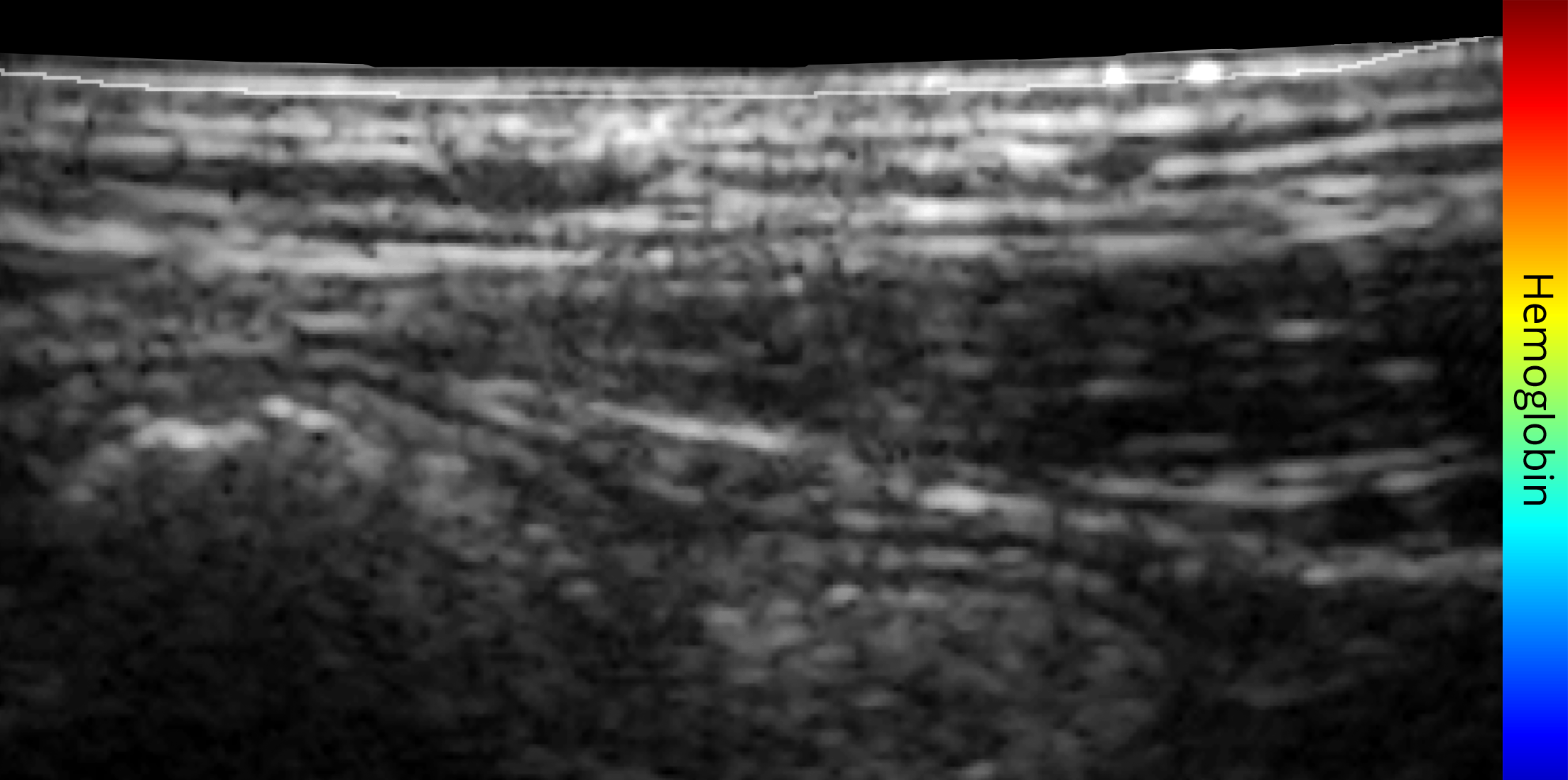
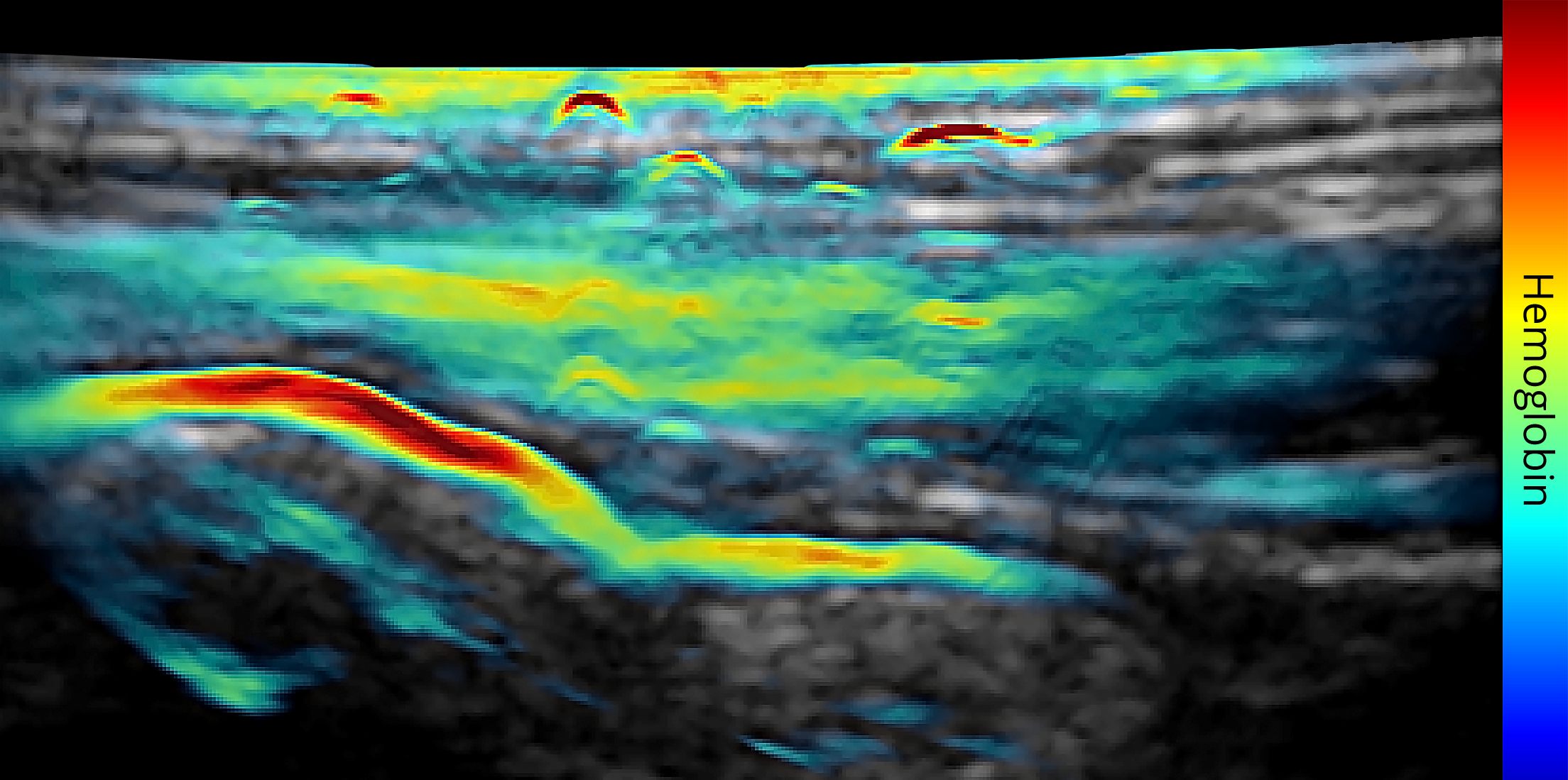
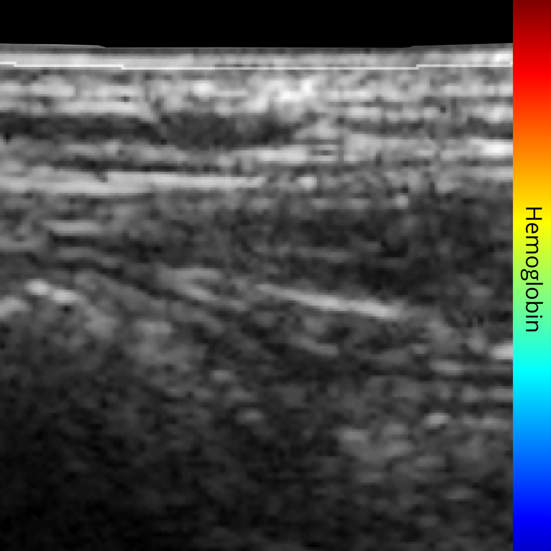
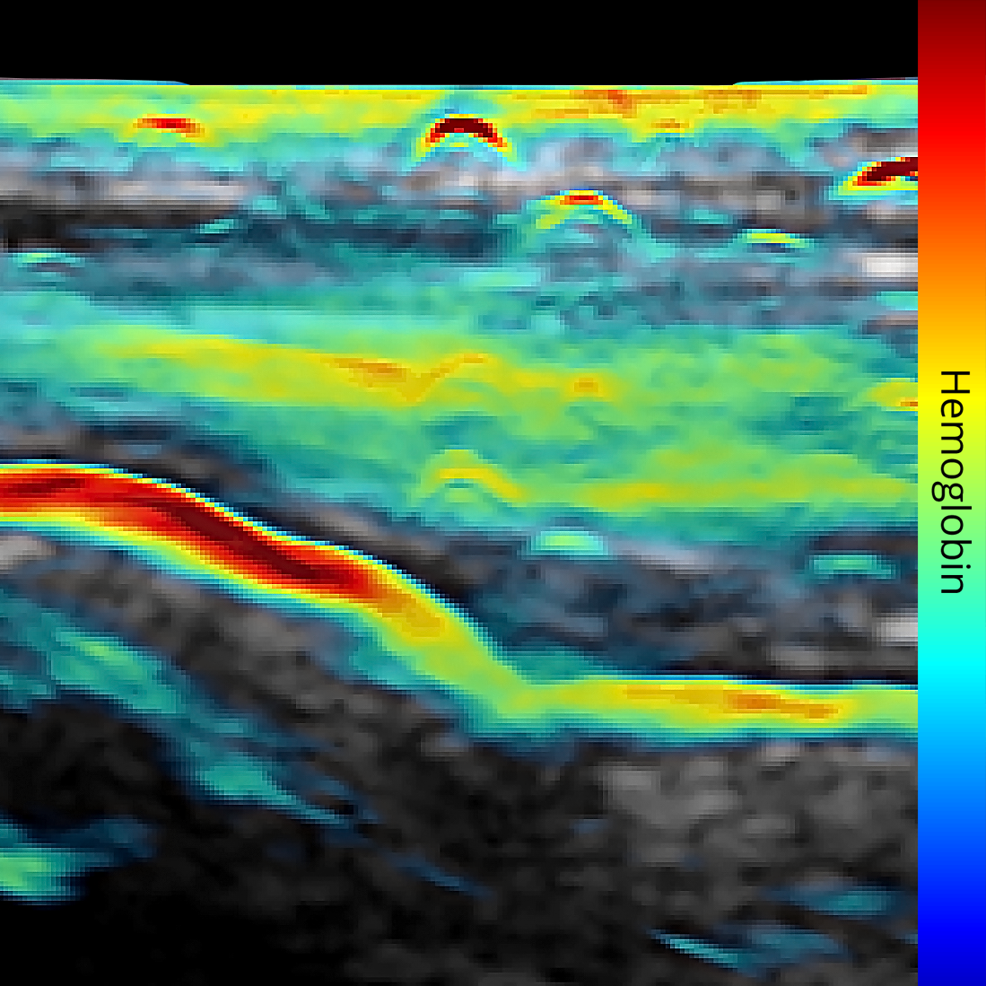
Bringing spectral information to greyscale ultrasound
During acquisition, both B-mode ultrasound and optoacoustic data are recorded. The optoacoustic signals are then spectrally analyzed and overlaid on the co-registered ultrasound data, providing information on specific disease biomarkers in addition to structural information.
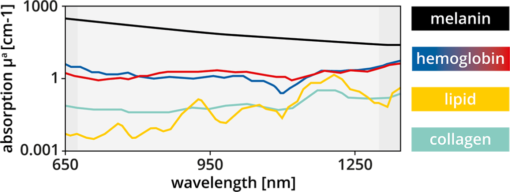
Multispectral analysis
Different biomolecules – such as hemoglobin, collagen, melanin, water, lipids – have different absorption properties at different wavelengths. By varying the wavelength with multispectral imaging, the signals from specific biomarkers can be isolated for visualization and quantification.