PRECLINICAL ORGAN DYSFUNCTION OF THE
DIGESTIVE SYSTEM
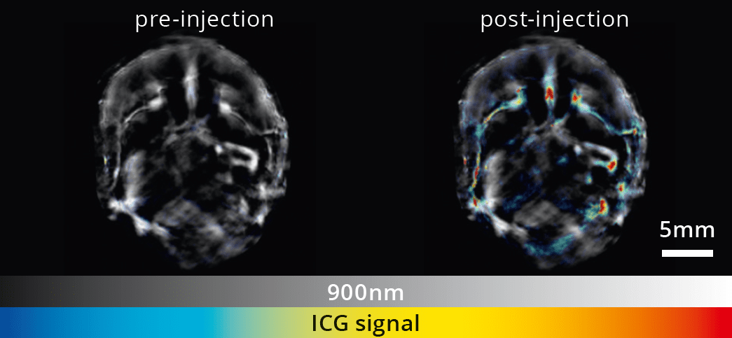
Therapeutic intervention of events caused by acute and chronic disease processes requires a detailed understanding of the mechanisms behind the injury. MSOT has been used to assess liver function in a range of liver injury models. The simple approach taken with MSOT can measure clearance of dyes like indocyanine green in real time to give a value of liver function that correlates well with traditional biomarkers. This approach allows non-invasive and easy monitoring of liver function through the full course of the disease process.
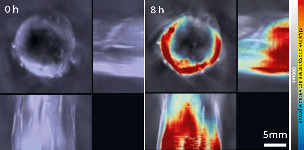
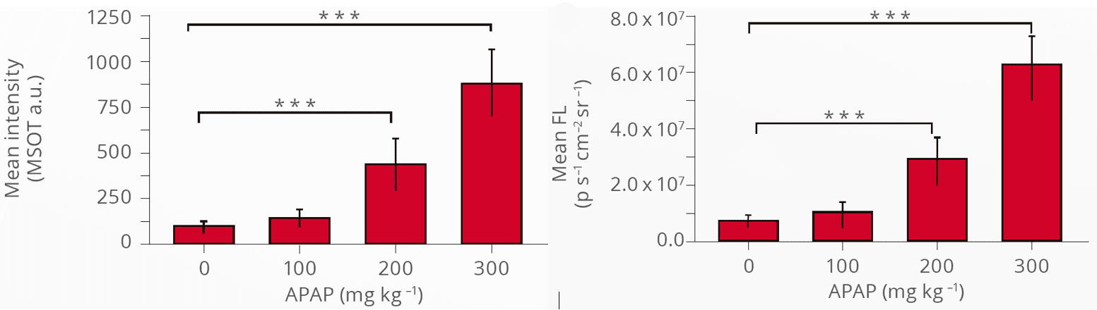
Single wavelength MSOT contrast in greyscale with an overlay of signal from the injected contrast agent. MSOT contrast agent signal was quantified at multiple timepoints and compared with fluorescence.
Assessment of liver injury via MSOT with an activatable contrast agent
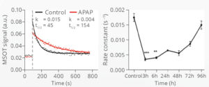
MSOT can be used to measure liver injury using a contrast agent, activated by hepatic alkaline phosphatase, before and after exposure to the hepatotoxicant acetaminophen (APAP). Increased alkaline phosphatase levels represent a biomarker of liver injury.
Wu et al. Nat Commun. 2018
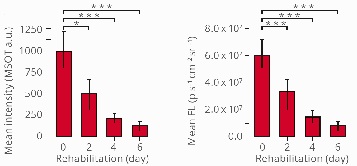
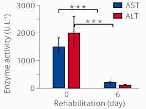
Alkaline phosphatase measurements with MSOT following therapy
Contrast agents can also be used to monitor liver recovery after therapy. The time course of MSOT measurements closely resembles measurements of fluorescence enzymatic activity in the blood.
Wu et al. Nat Commun. 2018
Quantification of MSOT and fluorescence signals as well as blood enzymatic activity in the days following therapeutic intervention.
Single wavelength MSOT signal in greyscale overlaid with spectrally unmixed ICG signal, which was quantified before, during and after ICG injection. This yielded pharmacokinetic parameters that could be measured repeatedly.
Impaired hepatic clearance of ICG quantified by MSOT
Longitudinal quantification of ICG, which is subject exclusively to hepatobiliary excretion, allows for the non-invasive assessment of liver function in mice with APAP-induced liver injury versus healthy control animals. Reduced liver function results in a delayed excretion of ICG from the organ.
Using several approaches, MSOT allows for the investigation of liver injury and hepatic regeneration.
Brillant et al. Toxicol Appl Pharmacol. 2017
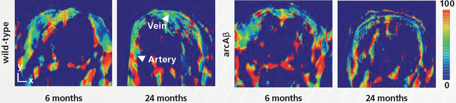
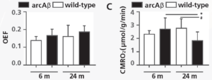
In vivo assessment of brain oxygenation in arcAβ mice using MSOT
Brain oxygen extraction fraction (OEF) remained unchanged in Alzheimer’s arcAβ mice, while the cerebral metabolic rate of oxygen (CMRO2) was reduced in Alzheimer’s mice at 24 months of age. MSOT can non-invasively assess vascular deficits related to neurodegeneration.
Ni et al. Photoacoustics. 2018
MSOT-derived oxygen saturation maps shown for wild-type and arcAβ mice at 6 and 24 months of life. MSOT and MRI measurements were combined to calculate brain hemodynamic parameters such as OEF and CMRO2.