PRECLINICAL NEUROLOGICAL DISORDERS OF THE NERVOUS SYSTEM

Alzheimer’s disease (AD) is a progressive neurodegenerative disorder that contributes to most dementia cases in adults. Preclinical models of AD aid in understanding the disease, and imaging plays a key part in this. Through MSOT imaging, it is possible to visualize plaque formation through the application of targeted molecular imaging agents and determine the change in both perfusion and oxygenation in neural tissue, thus providing a greater understanding of the disease progression and impact in vivo.

MSOT-derived oxygen saturation map of the brain as a jet overlay with transparency on an MRI measurement of the same animal.

Functional measurements co-registered with MRI

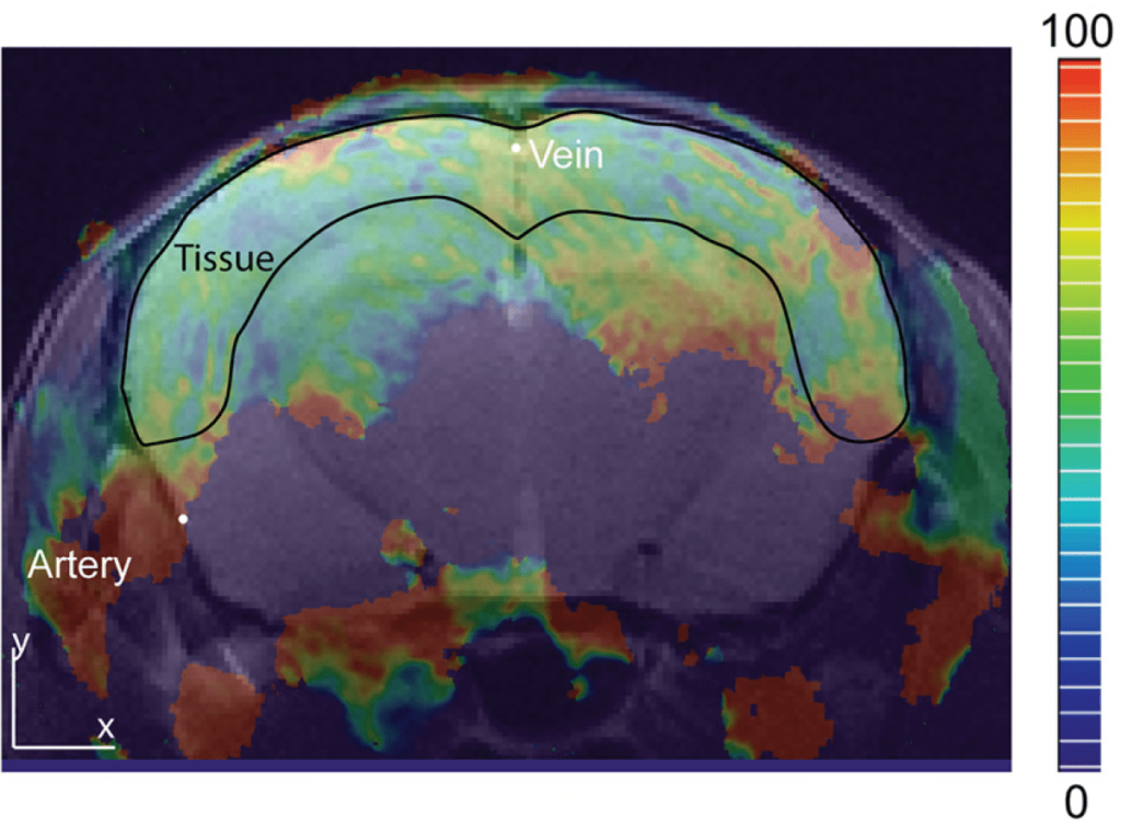
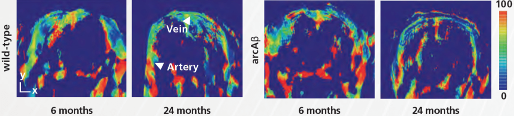
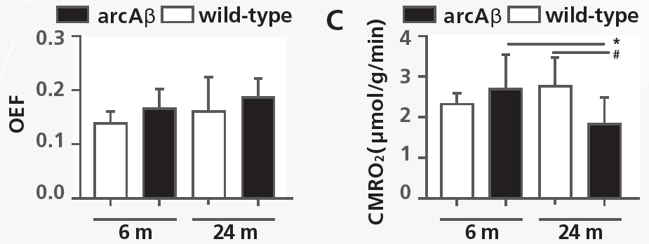
MSOT measurements can be co-registered with MRI. The combination of the two imaging modalities enables calculation of cerebral blood flow (CBF), oxygen saturation, brain oxygen extraction fraction and – in conjunction with perfusion imaging for CBF – the cerebral metabolic rate of oxygen (CMRO2), all key measures of brain hemodynamic function. 


Ni et al. Neurophotonics. 2018

In vivo assessment of brain oxygenation in arcAβ mice using MSOT

Brain oxygen extraction fraction (OEF) remained unchanged in Alzheimer’s arcAβ mice, while the cerebral metabolic rate of oxygen (CMRO2) was reduced in Alzheimer’s mice at 24 months of age. MSOT can non-invasively assess vascular deficits related to neurodegeneration. 


Ni et al. Neurophotonics. 2018

MSOT-derived oxygen saturation maps shown for wild-type and arcAβ mice at 6 and 24 months of life. MSOT and MRI measurements were combined to calculate brain hemodynamic parameters such as OEF and CMRO2.