PRECLINICAL METABOLIC DISORDERS
OF THE
INTEGUMENTARY SYSTEM
Metabolic disease research has intensified around the role of metabolically active brown adipose tissue (BAT), which has offered a therapeutic target in diabetes. Non-invasive pre-clinical imaging of BAT will enable longitudinal assessment of metabolic changes and may facilitate development of novel treatment therapies. Positron emission tomography (PET) / computed tomography (CT) is a routinely used molecular imaging technique to assess functional characterization of BAT activity, but suffers from relatively poor spatial resolution and requires the use of ionizing radiation. MSOT has shown promising results in non-invasive, label-free measurements of BAT metabolism and differentiation between diseased and healthy states. A multi-modal approach of using MSOT in combination with PET/ magnetic resonance imaging has demonstrated the potential to advance the preclinical screening of biomarkers that promote browning of the fat, with the possibility of translating these discoveries into clinical studies in humans.
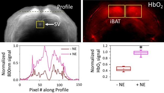
Brown adipose tissue, shown as single-wavelength (greyscale) or oxygenated hemoglobin (HbO2, red). Regions of interest highlight the Sulzer vein (SV) and areas of high brown adipose tissue (iBAT). MSOT signal quantified across a linear profile, and before and after norepinephrine activation.
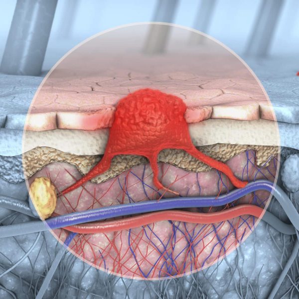
Brown adipose tissue imaging in mice as a biomarker of energy consumption
Brown adipose tissue and the Sulzer vein can be localized with MSOT based on endogenous contrast. MSOT can quantify brown fat metabolism through enhanced hemoglobin levels, as shown in studies with norepinephrine activation of this tissue.
Reber et al. Cell Metab. 2018
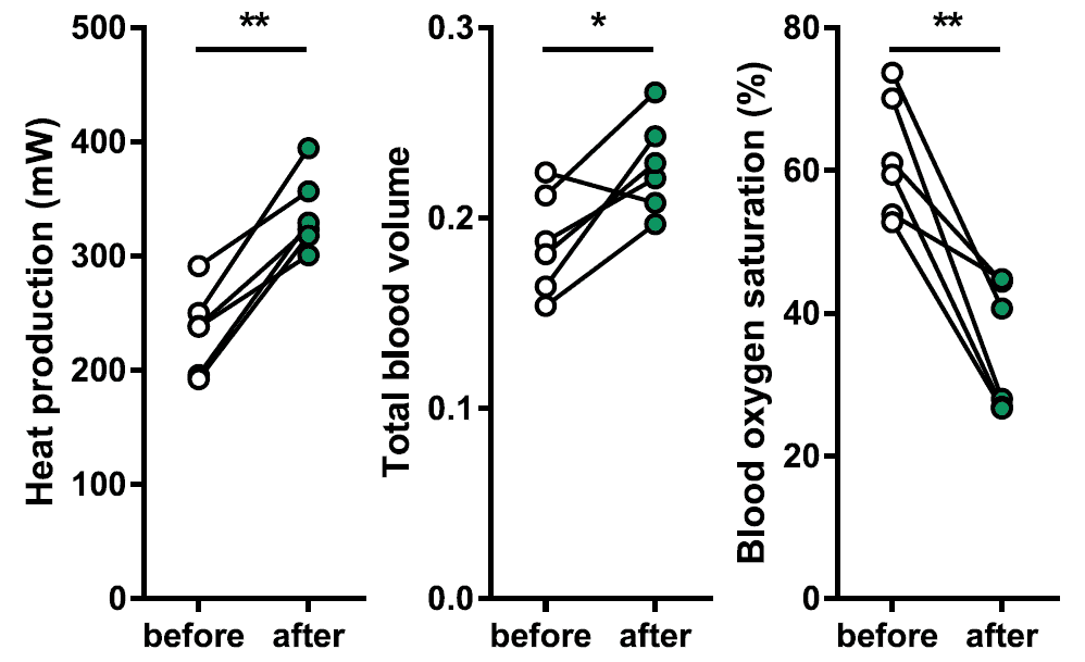
Comparison of MSOT measurements with indirect calorimetry
Heat production was measured by indirect calorimetry before and after injection of the thermogenic hormone secretin. MSOT measurements of total hemoglobin and oxygen saturation show increases and decreases (respectively), in line with calorimetric measurements.
Li et al. Cell. 2018
Heat measurement via indirect calorimetry (left), and quantification via MSOT of total blood volume (middle) and blood oxygen saturation (right).
Quantification of MSOT spectral unmixing of iRFP. MSOT signals increase over time after injection with a beta-adrenergic agonist.
Measurement of brown adipose tissue through MSOT detection of a genetic reporter
As an alternative to measuring vascular signals, browning of white adipose tissue in response to chemical or cold exposure in mice can be assessed via a genetic reporter system designed to monitor adipose browning. The reporter iRFP absorbs light in the near infrared range and can then be imaged by MSOT, revealing molecular changes in adipose tissue over time.