PRECLINICAL METABOLIC DISORDERS
OF THE
CIRCULATORY SYSTEM
Preclinical research in murine models is essential to study immunometabolic processes and molecular mechanisms driving circulatory disease such as atherosclerosis. It is critical to identify atherosclerosis in its early stage, and a non-invasive approach that can provide high spatial and temporal resolution of anatomical and hemodynamic information is necessary to accurately diagnose disease and assess therapy. Conventional imaging techniques such as magnetic resonance imaging and computed tomography suffer from poor sensitivity for signal detection, while single photon emission computed tomography and positron emission tomography suffer from poor spatial resolution. Optoacoustic imaging with novel nanoprobes has shown the ability to provide high contrast-enhanced imaging of atherosclerotic plaques in a living mouse model of atherosclerosis.
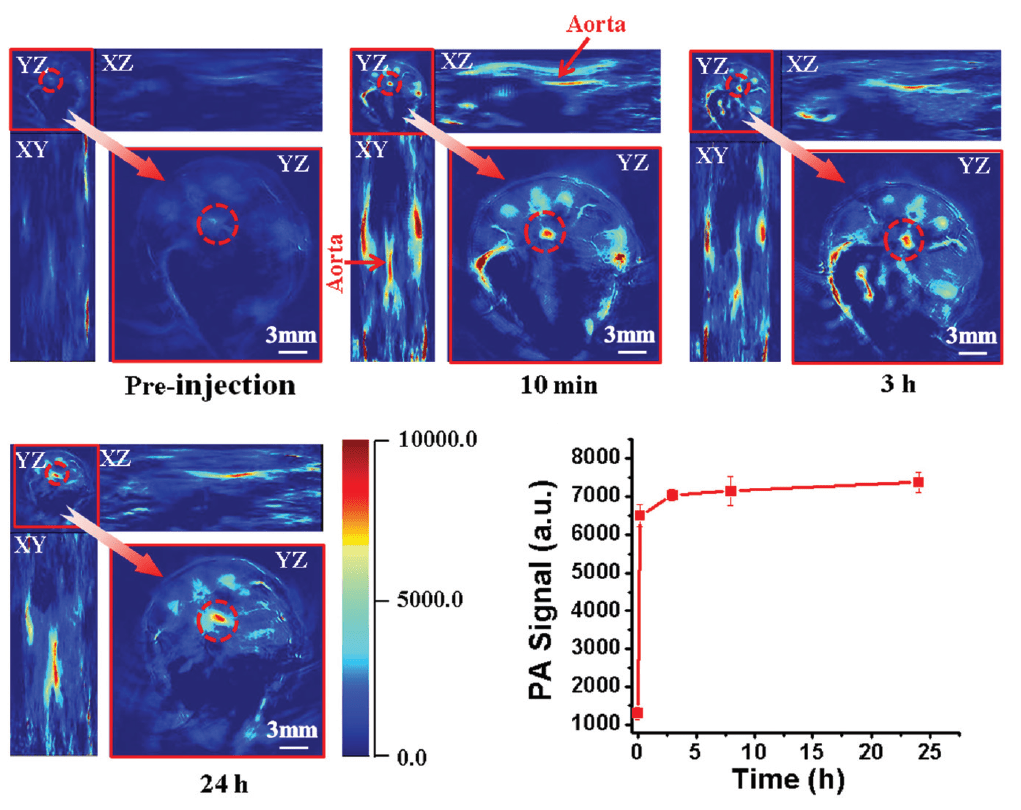
Time course of MSOT imaging of ApoE−/− mice after intravenous injection of ICG@PEG-Ag2S.
Atherosclerotic plaques visualized with specific nanoprobes
Apolipoprotein E knockout (ApoE−/−) mice were injected with ICG@PEG-Ag2S, a probe that binds to atherosclerotic plaques and can be visualized with MSOT. Enhanced MSOT signal was seen during the 24 hours following injection, with uptake consistent with ex vivo determination of plaque position.