PRECLINICAL INFLAMMATION
OF THE
INTEGUMENTARY SYSTEM
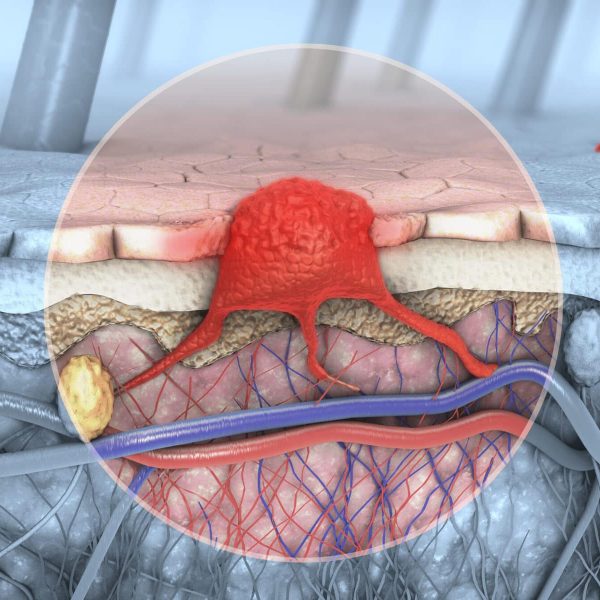
Cutaneous autoimmune diseases like Scleroderma can cause inflammation in the skin and other areas of the body, leading to patches of thickened skin and injury of small vessels. Current diagnosis and monitoring is conducted mainly via visual inspection of the skin or dermatoscopy, which only allows for a subjective evaluation of the skin surface. In a preclinical study, raster-scanning optoacoustic mesoscopy (RSOM) was proven to enable high-resolution quantitative assessment of Scleroderma in mouse ears, such as changes in skin vascularization including blood volume and hemoglobin content, with the potential to enhance understanding of the disease status and therapy response.
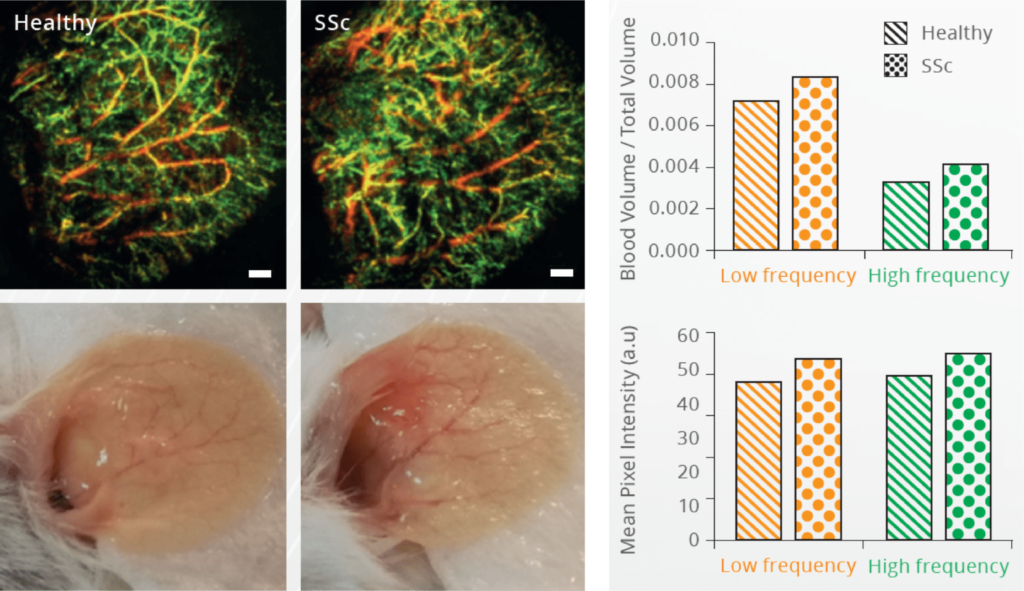
Before and after treatment. RSOM images and photographs of the ear in a healthy mouse and a mouse with systemic scleroderma (SSc), and quantification of the RSOM images.
Detection of SSc in the mouse ear using RSOM
The ear can be imaged by RSOM with minimal animal preparation. A slight increase in blood volume and higher mean pixel intensity could be detected with RSOM in the ear of animals with systemic scleroderma (SSc), suggesting that a detection of the systemic condition is possible throughout different body parts.
Courtesy of Galapagos SASU in Romainville, France
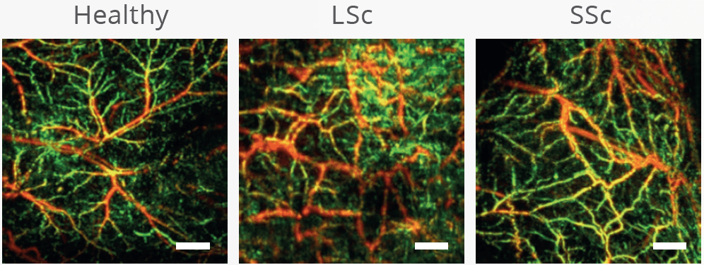
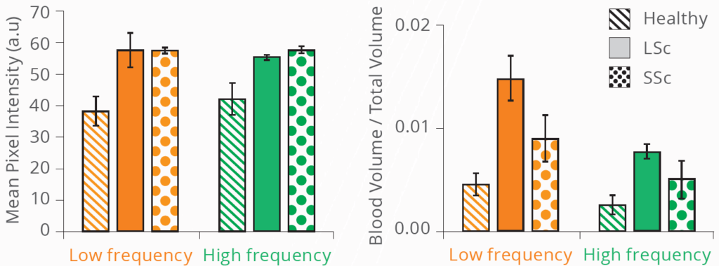
Assessment of LSc and SSc in the skin of mice using RSOM
The localized (non-systemic) form of scleroderma (LSc) affects the skin of only the face, hands, and feet while the more severe systemic form (SSc) also affects visceral organs. RSOM offers a unique tool to monitor progression and regression of the disease state. Both types of Sc show a higher mean pixel intensity as well as an increased blood volume in the RSOM images compared to the skin of healthy animals. LSc shows a significantly higher blood volume compared to SSc.
Courtesy of Galapagos SASU in Romainville, France
RSOM images of healthy and localized or and systemic forms of scleroderma, and the associated quantification of vascular structures.
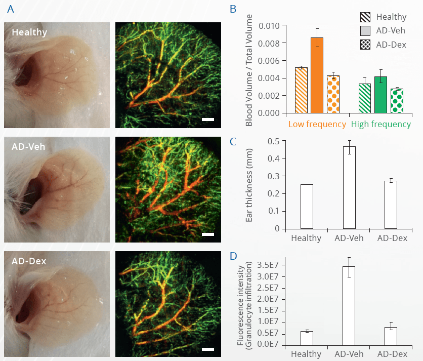
Photographs of mouse ears with and without dermatitis, and with treatment, compared with high resolution vascular RSOM images. Large (low frequency) and small (high frequency) structures are color-coded. RSOM signals are quantified for each group.
RSOM measurements of the ears in mice with dermatitis after treatment
Mice with atopic dermatitis (AD) showed an increased blood volume noninvasively measured by RSOM. Measurements of the ear thickness and granulocyte infiltration into the ears correlated well with the RSOM results. However, RSOM has the benefit of detecting differences in single vessels non-invasively over time, without the need to inject any additional contrast agents or to conduct histology.