The current gold standard of diagnosis of inflammatory bowel diseases (IBD) includes invasive procedures such as colonoscopy and biopsies, which carry risk to the patients. A non-invasive imaging modality that can be used to both accurately identify early inflammatory changes in the small bowel and colon as well as longitudinally monitor the status of disease will allow better understanding of the disease and treatment efficacy. Due to its ability to detect deep tissue oxygenation without the need of an exogenous contrast agent, MSOT has proved to be a promising imaging modality that effectively identifies colitis – one of the most prevalent types of IBD – in a murine model. MSOT has demonstrated diagnostic accuracy, with good correlation with the current gold standards of colonoscopy and tissue histology.
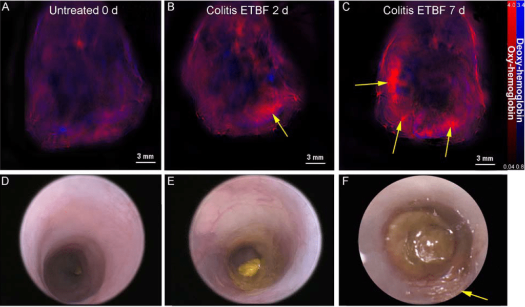
Oxy- and deoxy-hemoglobin MSOT contrast before and at two time points following induction of colitis, compared with endoscopy at those same timepoints.
Multispectral optoacoustic tomography depicts inflammatory changes in murine colitis
Mice were treated with clindamycin and streptomycin for 4 days followed by administration of enterotoxic Bacteroides fragiles via oral gavage in order to induce colitis. Mice were then scanned by MSOT, and the non-invasive oxygenation images were compared to endoscopy.
Bhutiani et al. J Nucl Med. 2017
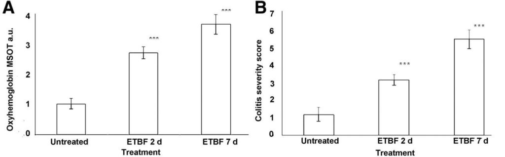
Quantification of inflammation in a mouse model of colitis
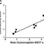
MSOT correlates well with endoscopic scores, showing increased signal with higher colitis severity scores. MSOT could potentially be used to noninvasively visualize vascularity in the gut in inflammatory diseases.
Bhutiani et al. J Nucl Med. 2017
Mean MSOT signal intensity for oxy-hemoglobin correlates for untreated mice compared treatment groups, and with the Murine Endoscopic Index of Colitis Severity.