PRECLINICAL INFLAMMATION
OF THE
CIRCULATORY SYSTEM
Studying animal models of myocardial infarction (MI) plays an important role in the diagnosis, prevention, and therapeutic evaluation in human patients. While promising, in vivo optical imaging in near-infrared (NIR) spectrum lacks spatial resolution and overall accuracy at increased penetration depths due to light scattering. MSOT overcomes these limitations and has demonstrated selective targeting with NIR optical contrast agents in a murine MI model. Together with targeted agents, MSOT can help better understand biological processes during myocardial healing in vivo and in evaluating novel therapeutic strategies.
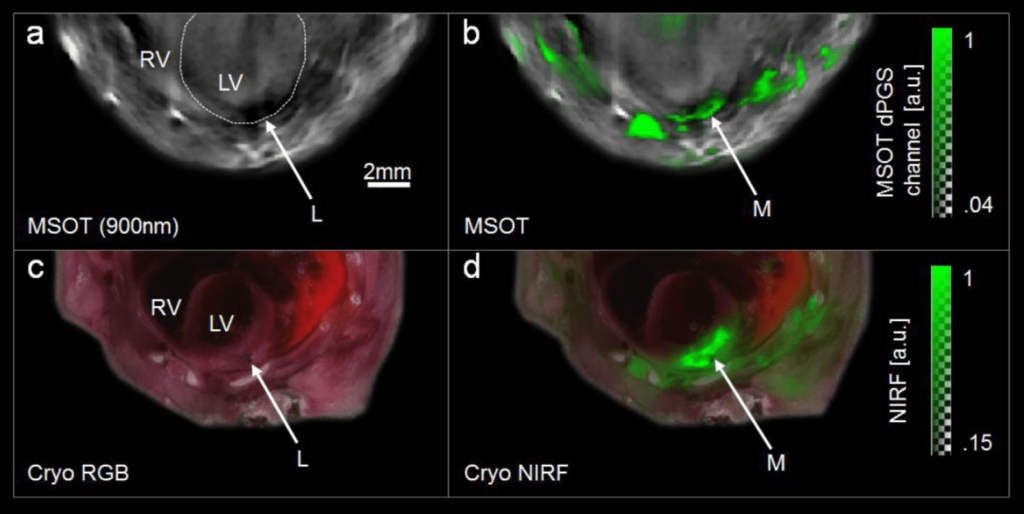
Single wavelength MSOT contrast is shown in greyscale, with an overlay of selectin signal in green. Color images show frozen mouse sections with fluorescence overlay.
MSOT contrast enhancement in myocardial infarction
MSOT was used to noninvasively image MI in a murine model ligating the left anterior descending artery. Dendritic polyglycerol sulfates (dPGS), which have specificity for P and L selectins, were injected two days post-occlusion. Inflammatory processes that occur during cardiac injury can be visualized by MSOT.