PRECLINICAL FIBROSIS OF THE
MUSCULOSKELETAL SYSTEM
Lethal muscular diseases like Duchenne Muscular Dystrophy (DMD) pose significant burden not only on patients, but also their families and the healthcare system. The current status of severity and therapy assessment relies on functional tests involving physical challenges, which are highly variable and unsuitable for young infants and other non-ambulant patients. Optoacoustic imaging could offer an objective and quantitative tool to monitor the status of such patients. A translational study in piglets could show increased collagen content in the distal extremities of the DMD group, allowing for an easy and objective longitudinal assessment of disease progression and treatment outcome.
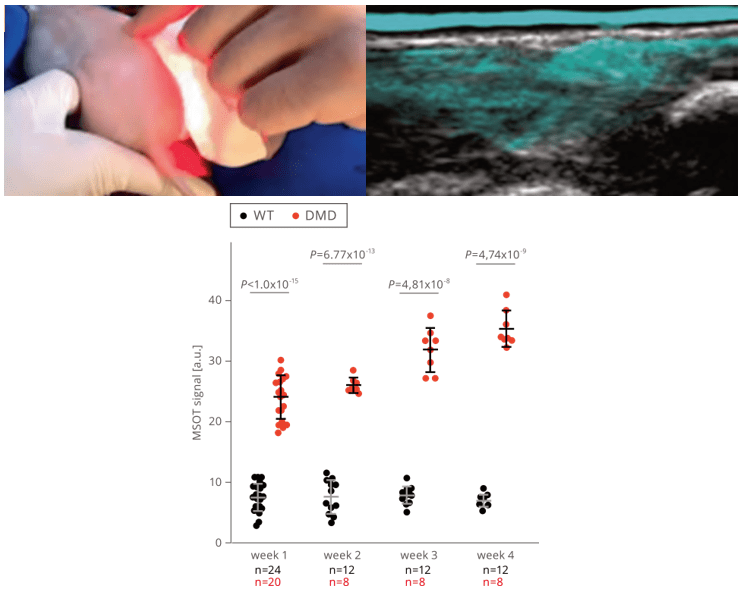
Longitudinal assessment of collagen content and ex vivo verification in DMD and WT piglets
The collagen content of the distal extremities was monitored non-invasively by MSOT imaging without the need for sedation, in both healthy wild-type (WT) and DMD piglets over 4 weeks. The results reveal an excellent sensitivity and specificity of MSOT-derived collagen signal detection to distinguish between healthy and diseased tissue from birth.
Regensburger et al. Nat Med. 2019
DMD and WT piglets scanned by MSOT with spectral unmixing for collagen. Collagen content in the muscle shown as cyan overlaid onto an ultrasound image.
Tissue staining for Sirius red and Masson trichrome used to visualize collagen as a biomarker of fibrosis.
Histological validation of MSOT measurements of fibrosis
Validation of the presence of fibrosis biomarkers was performed by immunohistochemistry. An increase of different types of collagens could be found in all DMD piglets present from week one of life, which strongly correlated with the MSOT findings.