Breast cancer is the most common invasive cancer in women. In addition to surgery, there are multiple approaches to therapy, including hormonal, chemo- and immuno-therapy as well as radiation therapy. Developing these therapies often begins in animal models, which can recapitulate many aspects of the disease. MSOT has been used in mouse models to differentiate cancer types based on vascularity, to elucidate therapeutic targets, and to test a variety of nanotheranostic agents.
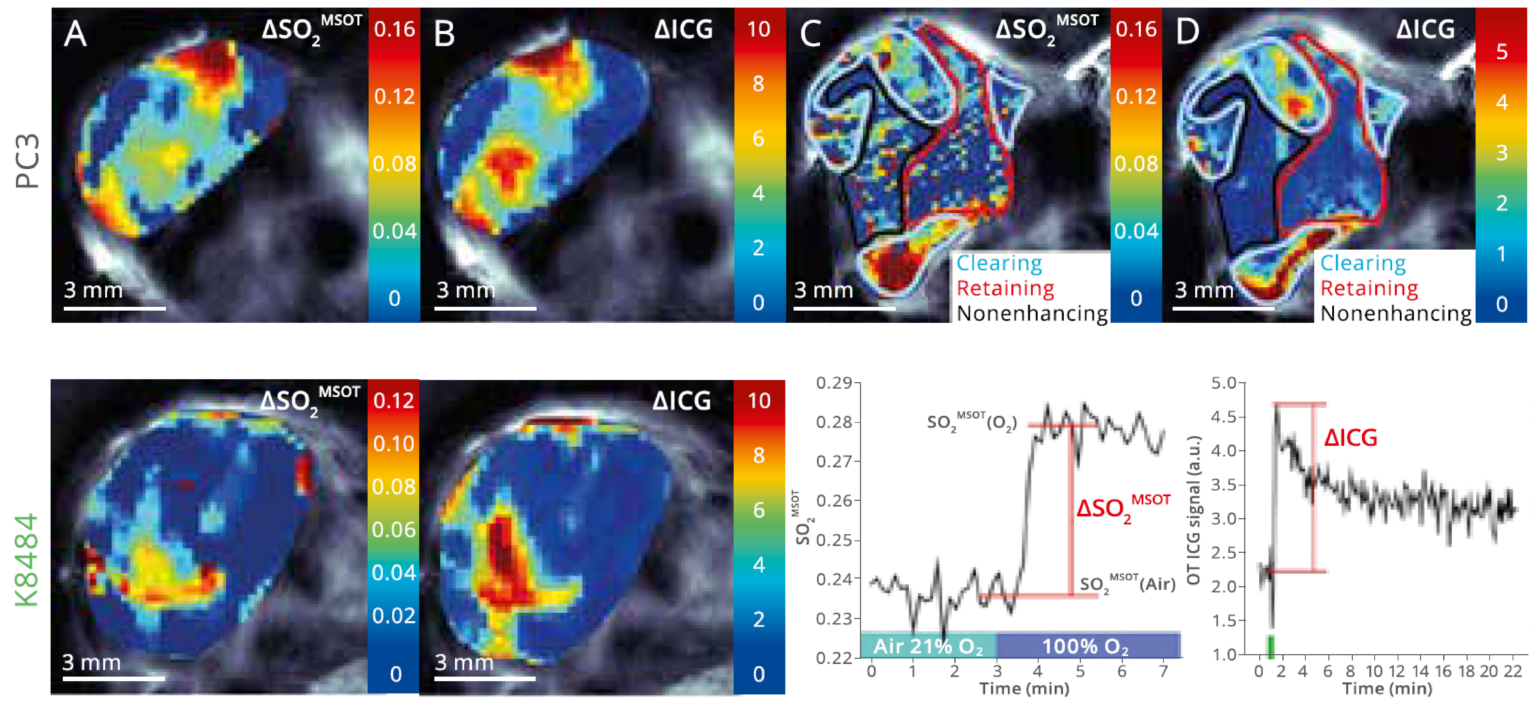
Comparison of oxygen enhanced (OE) and dynamic contrast enhanced (DCE) MSOT and DCE-MSOT in mice bearing tumors from a prostrate cell line.
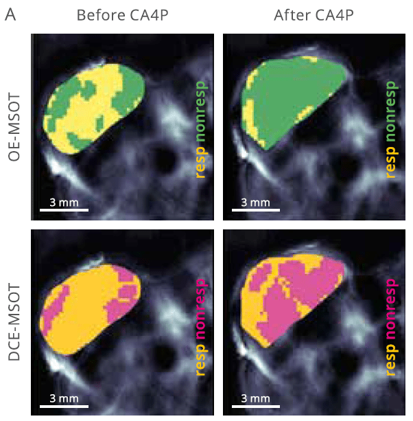
Oxygen challenges reveal tumor heterogeneity
Dynamic imaging is useful for determining vascular function in regions of interest. Oxygen-enhanced (OE) MSOT is a label-free, safe, and clinically translatable method designed to highlight tissue regions responsive to changes in oxygen concentration, as was demonstrated in this prostate cancer cell line. Dynamic contrast enhanced (DCE) MSOT utilizes injectable contrast such as indocyanine green (ICG) to identify regions with differential perfusion of contrast.
Tomaszewski et al. Cancer Res. 2018
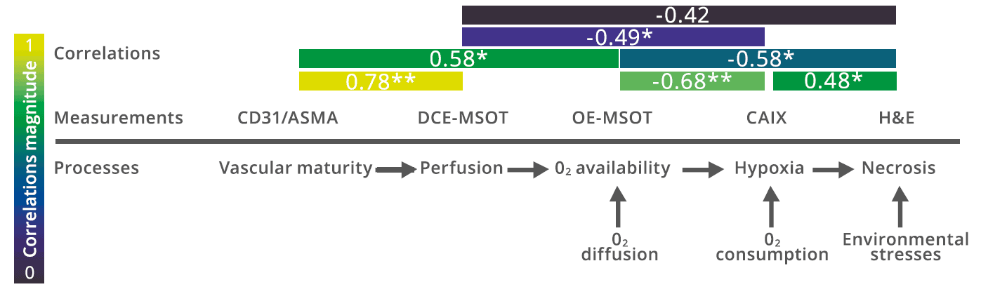
Comparison of functional MSOT assessments to histological gold standards
OE-MSOT signal is driven predominantly by tumor hypoxia and necrosis, whereas DCE-MSOT signal most strongly reflects vascular maturity as it relates to perfusion. A combination of the two MSOT biomarkers can reveal a unique and easily obtained perspective into the tumor vascular microenvironment.
Tomaszewski et al. Cancer Res. 2018
Correlation coefficients are shown between MSOT measurements—either ΔSO2 from OE-MSOT or ΔICG from DCE-MSOT—and histological measurements.
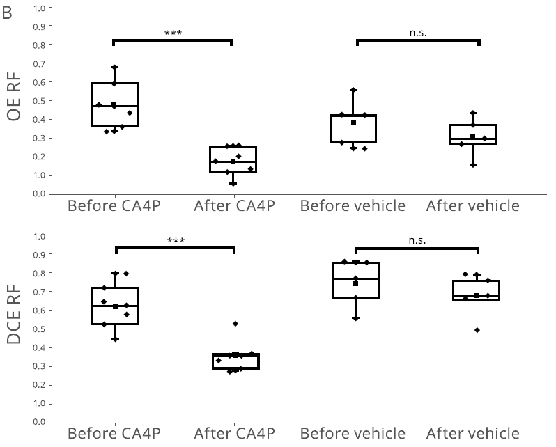
OE- and DCE-MSOT studies were used to generate a map of the responding fraction (RF) of the tumor, which can then be quantified before and after vascular therapy.
Tumor vascular response to therapy
Treatment with a vascular disrupting agent causes a massive vascular shutdown that can be measured non-invasively with MSOT through derivation of DCE-RF, which quantifies perfusion of an injected agent into the tumor. As an alternative to an injected contrast agent, hemoglobin-based OE-RF measurements, which measures changes in tumor oxygenation, are also sensitive to treatment.
Tomaszewski et al. Cancer Res. 2018
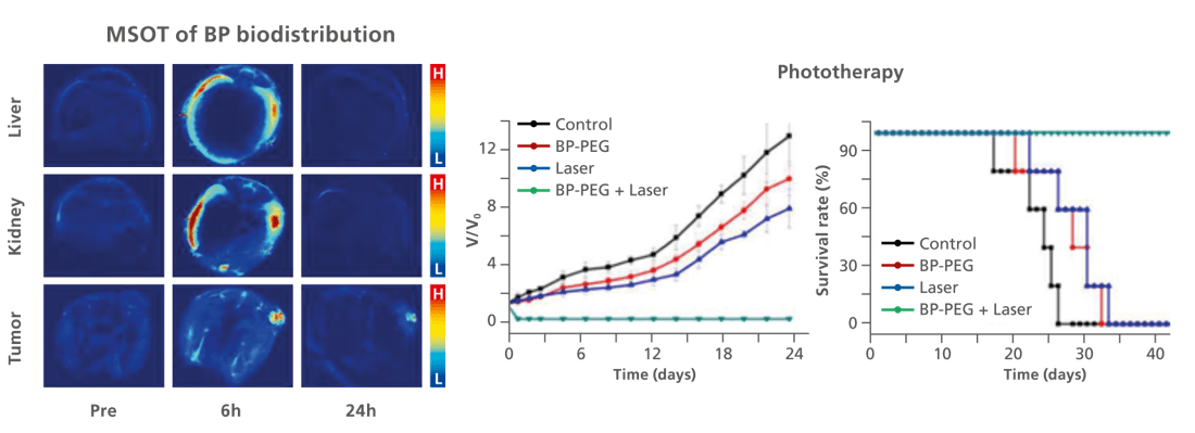
MSOT imaging as a guide for phototherapy using nanoprobes
MSOT was used to track the distribution of black phosphorous (BP) nanoparticles, selected for their theranostic potential of providing both MSOT contrast and photothermal therapeutic action. MSOT was used to determine the optimal time point for high tumor accumulation with low off-target binding, allowing successful use of phototherapy.
Sun et al. Biomaterials. 2016
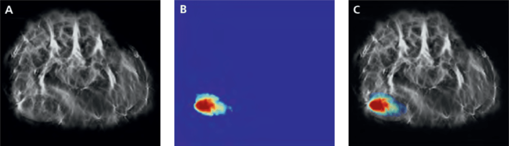
Spectrally unmixed nanoprobe signal before and after injection, showing peak tumor accumulation of the phototherapeutic agent.
Orthotopic mammary tumors implanted in mice, with single-wavelength contrast in greyscale and iRFP signal in jet.
iRFP fluorescent protein as a genetic reporter for tumor imaging
Thanks to its high spatial resolution and its ability to detect contrast agents at depth, MSOT can be used to track iRFP expression and tumor development in vivo.