Spread of cancer through the lymphatic vessels is usually a precursor to secondary distant tumor formation. Identifying and characterizing this spreading tumor tissue is key to understanding the metastasis process. In cases involving darkly absorbing melanoma cells, MSOT has been utilized to image the spreading of incredibly small numbers of cells within the lymphatic network. Furthermore, molecular imaging strategies have been employed with MSOT to identify spreading cells and characterize the tumor microenvironment.
Orthogonal maximum intensity projections of the legs and lymph nodes of control animals or mice bearing tumors in the hindlimb paw. Single wavelength MSOT contrast is shown in greyscale, and the activatable probe is shown as a jet overlay.
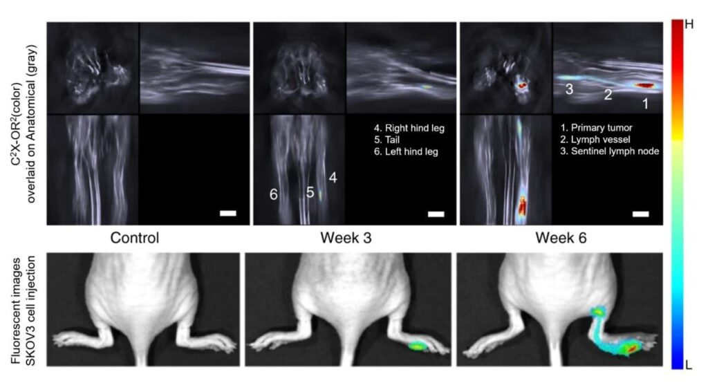
Detection of tumor metastasis with β-Gal sensitive probes
SKOV-3 ovarian cancer cells overexpressing β-Gal were injected in the right foot pad and allowed to metastasize to the popliteal lymph node. Following intratumoral injection of the xanthene-based optoacoustic probe C2X-OR2 to label β-Gal-containing cells, the primary and the metastatic sites were detected by means of MSOT and fluorescence imaging.
Wu et al. Nat Commun. 2018
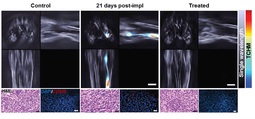
Detection of tumor metastasis with activatable probes
Overexpression of hyaluronidase in 4T1 breast primary tumor & metastatic cells activates an injected nanoprobe, resulting in spectral shift & signal enhancement. MSOT measurements are sensitive to subsequent cyclophosphamide treatment.
Wu et al. Adv. Funct. Mater. 2019
Orthogonal maximum intensity projections of the legs and lymph nodes of control animals or mice bearing tumors in the hindlimb paw. Single wavelength MSOT contrast is shown in greyscale, and the activatable probe is shown as a jet overlay.