Tumors in deep tissue in the colon or pancreas are usually managed by invasive resections. To make those resections as effective and small as possible, suitable imaging modalities are needed to help localize and demarcate those tumors. In several preclinical studies, it was shown that optoacoustic imaging could help to localize those deeper situated tumors via high resolution 3D views and the application of targeted contrast agents with high specificity before surgery. Furthermore, detailed vascular changes in such tumors could be monitored non-invasively with RSOM, helping to understand the progression and treatment success for cancer in the digestive system.
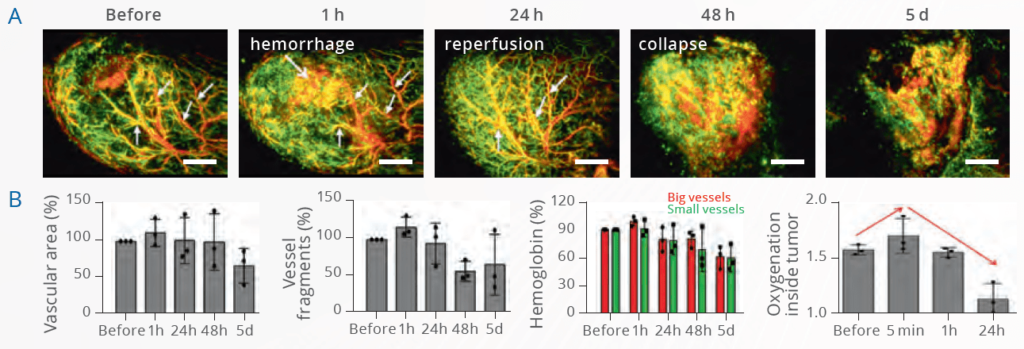
Long-term RSOM imaging of VTP effects in CT26 xenografts
RSOM is capable of imaging colon carcinoma xenograft vasculature on a microvascular level, revealing unprecedented insights into vascular-targeted photodynamic therapy (VTP). Upon localized light activation, an administered photosensitizer generates oxygen radicals within the tumor, leading to immediate occlusion of certain vessels, followed by hemorrhage. Initially non-vascularized areas appear with normalized vessels – indicating ischemic reperfusion – before the entire vasculature collapses. Multispectral RSOM, which distinguishes oxy- from deoxyhemoglobin, can reveal oxygenation changes.
Haedicke et al. Nat Biomed Eng. 2020
RSOM images of CT26 xenografts before and up to 5 days post-VTP therapy. RSOM images are quantified to provide metrics such as vascular area, vessel fragmentation, hemoglobin content, and oxygenation.
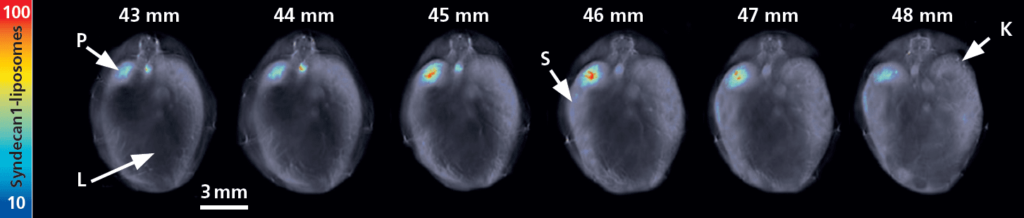
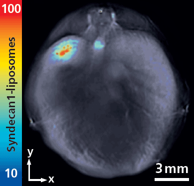
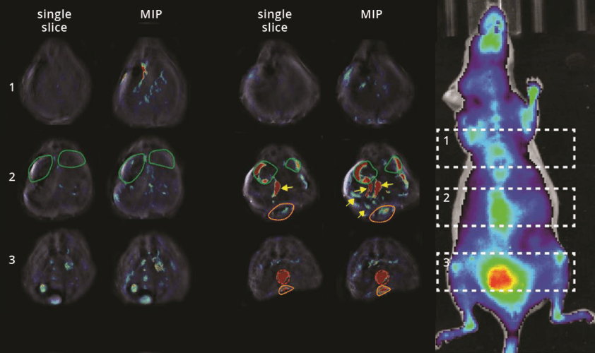
MSOT images of different positions of an orthotopic pancreas tumor and a maximum intensity projection of those positions. Single-wavelength contrast shown in grayscale, and spectrally unmixed signal from the peptide ligand of Syndecan-1 shown as a jet overlay.
Orthotopic pancreas tumor targeting with a peptide-based ligand, Syndecan-1
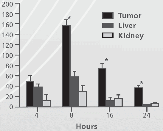
MSOT can be used with custom-made contrast agents. For example, a peptide ligand specific for Syndecan-1, which is a protein associated with avb3 integrin expression as well as cellular proliferation, can be visualized by MSOT to quantify biodistribution and tumor accumulation in an orthotopic pancreas tumor model.
Yin et al. J Nanobiotechnology. 2015
MSOT assessment of orthotopic pancreatic tumor targeting with cRGD-800CW-TCO
Developing probes that bind to biomarkers expressed solely in a disease state opens a wide range of applications that improve molecular diagnosis and potential for therapy. In cancer, targeting overexpressed biomarkers can enable improved delivery of chemotherapeutics or enhance radiotherapy. By using MSOT in this fashion, it is possible to differentiate primary tumors and small, deep-seated metastasis from clearance organs.
Napp et al. Int J Cancer. 2018
Single MSOT cross-sections or several cross-sections shown as a maximum intensity projection. Single-wavelength contrast shown in grayscale, and MSOT contrast from the peptide ligand shown as a jet overlay. MSOT compared to planar fluorescence.