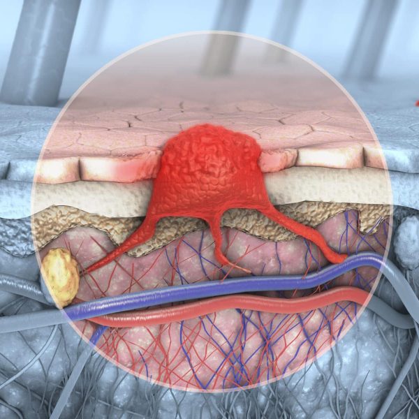
CLINICAL METABOLIC DISORDERS OF THE
INTEGUMENTARY SYSTEM
Although the process has historically been challenging, measurement and imaging of metabolic activity has great relevance to disease conditions like obesity and diabetes. The modulation of metabolic activity by pharmacological means is seen as a counter to these disease conditions, and the ability to assess local metabolic activity with MSOT is a valuable tool in this assessment. As well as the capability to visualize and quantify activation of brown adipose tissue through alternation in local blood oxygenation, it is also possible to morphologically directly discriminate between brown and white adipose tissue in vivo using MSOT.
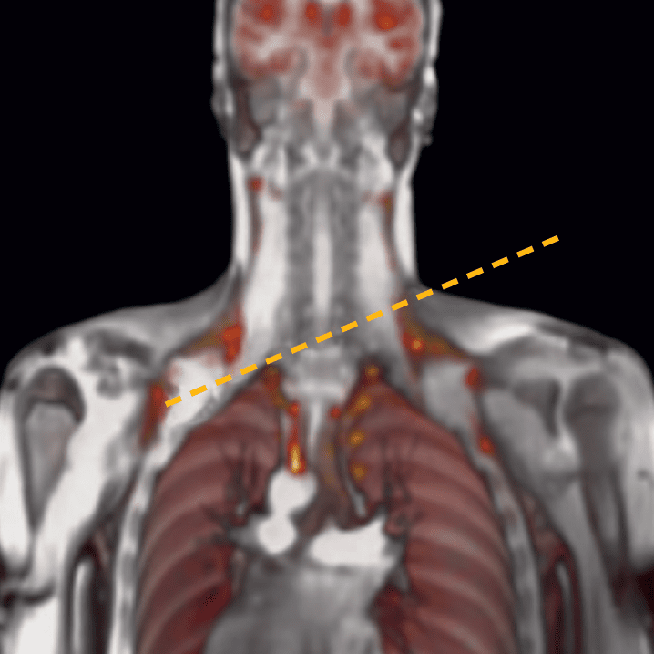
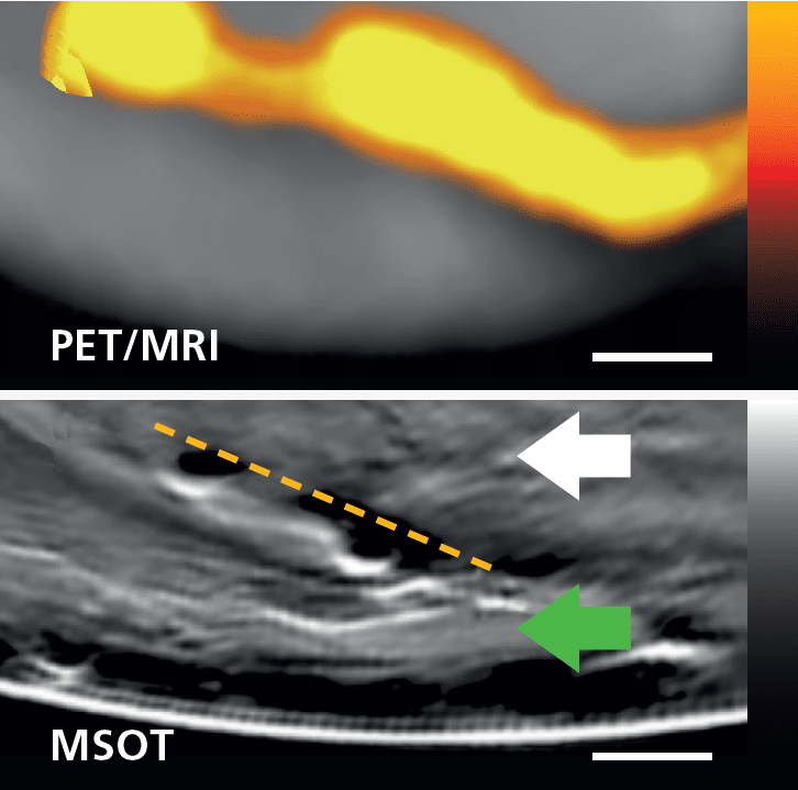
MSOT imaging of BAT in the upper torso, compared to imaging with PET/MRI using 18F-FDG.
Brown adipose tissue imaging as a surrogate for metabolic activity
Brown adipose tissue (BAT) can be measured directly by MSOT and compared to MRI-PET of the same region. MSOT measurements correlate with indirect calorimetry, thus demonstrating that MSOT can potentially visualize brown fat in different areas of the body and quantify metabolic activity in real-time.
Reber et al. Cell Metab. 2018
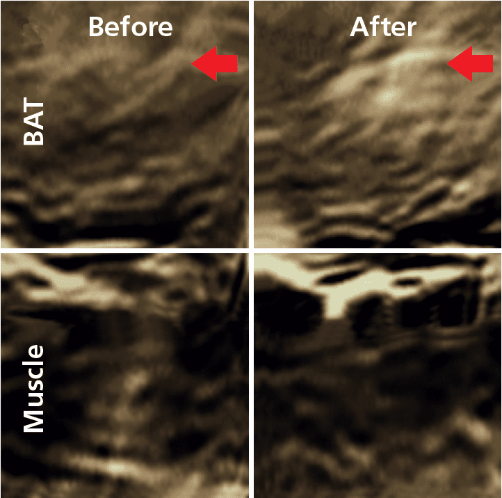
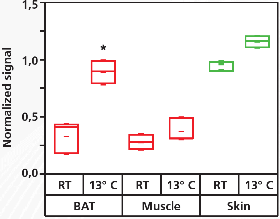
MSOT measurement of BAT activation
Brown adipose tissue (BAT) activation can be induced via cold exposure of the whole body by means of a suit perfused with 13˚C cold water. In a clinical pilot study with MSOT, BAT and muscle was imaged before and 10 minutes after cold activation. In BAT, the oxygenated hemoglobin (HbO2) intensity increased significantly following cold activation, whereas no significant change was seen in muscle.
Reber et al. Cell Metab. 2018
Quantification of MSOT signals before and after cold stimulation in BAT, muscle, and skin.