Methods for imaging vascular structures include ultrasound (US), magnetic resonance (MRI), and X-ray computed tomography (XCT). US is non-invasive but suffers from an inherent lack of sensitivity and resolution. MRI and XCT provide high resolution but require injection of contrast agents. In early clinical trials, MSOT has demonstrated the potential to provide high-resolution imaging of vascular morphology and function without exogenous contrast, at different scales for superficial microvasculature as well as deeper-lying vessels.
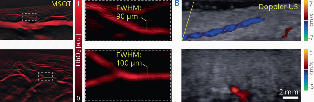
MSOT imaging of venules and arterioles in a healthy volunteer, compared with Doppler ultrasonography (Doppler US) of the same field of view.
Optoacoustic imaging can detect vasculature beyond the sensitivity of Doppler ultrasonography
MSOT can resolve both major vascular structures as well as those less than 90 µm in diameter, depending on the system configuration. Spectral unmixing further allows for relative quantification of endogenous molecules such as oxyhemoglobin (HbO2), deoxyhemoglobin (Hb), and oxygen saturation (sO2 MSOT) in vessels and tissues. The high spatiotemporal resolution of MSOT also provides the ability to observe real-time vascular physiological events, such as arterial pulse.
Taruttis et al. Radiology. 2016
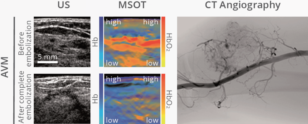
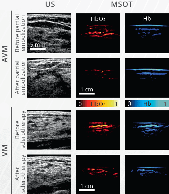
Optoacoustic imaging can detect changes after vascular intervention
Differential diagnosis and treatment of vascular abnormalities such as venous and arteriovenous malformations (VM and AVM, respectively) are often challenging, with a high rate of misdiagnosis and difficulties with determining therapeutic efficacy. The molecular information provided by MSOT is valuable for immediate evaluation of response to therapy.
Masthoff et al. JAMA Dermatol. 2018
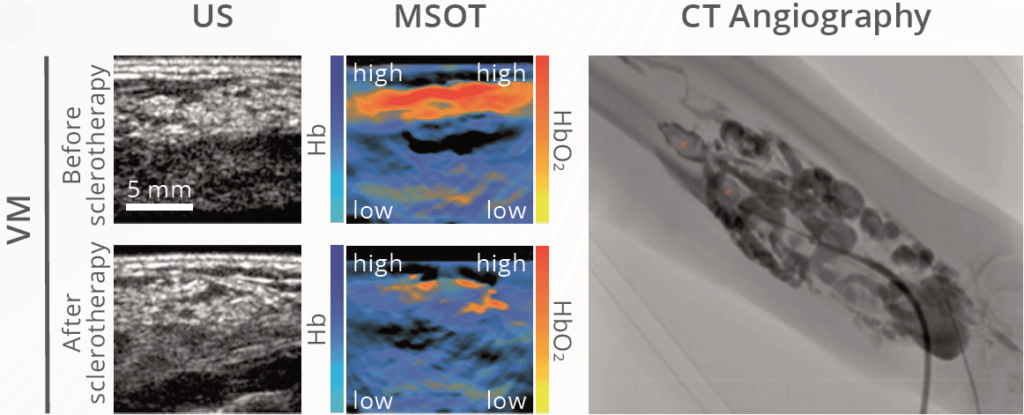
Evaluation of vascular malformations before and after therapy using MSOT. Ultrasound is compared to spectrally unmixed oxygenated hemoglobin (yellow and red scale) and deoxygenated hemoglobin (blue scale), with CT angiography shown as a reference.
Spectrally unmixed MSOT signals show oxygenated hemoglobin (yellow and red scale) and deoxygenated hemoglobin (blue scale). Ratios of MSOT signals are quantified before and after embolotherapy. Ultrasound signal shown in grayscale.
Quantification of therapeutic efficacy in AVM and VM using MSOT
Using MSOT, the mean ratio of spectrally unmixed HbO2 and Hb signal can be used as an objective measure of therapeutic response in AVM and VM. Following therapy, clear qualitative differences can be observed in MSOT HbO2 and Hb signal in both AVM and VM patients studied. Quantitative results showed significant reduction of the mean HbO2:Hb ratio in AVM and VM patients, validated by standard MRI or angiography.