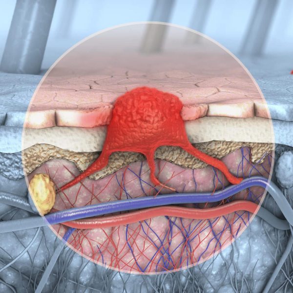
Inflammatory skin diseases span a wide field, including psoriasis, eczema, Rosacea, and dermatitis. The established method of diagnosis is visual skin inspection via dermatoscopy. While this allows for a detailed view of the epidermis, it does not allow for an assessment of disease progression in deeper skin layers. High-resolution optoacoustic imaging provided by RSOM (raster-scanning optoacoustic mesoscopy) has shown the potential to more accurately quantify disease status at depth by looking at parameters such as hemoglobin concentration, vessel diameter, and vessel bifurcation.
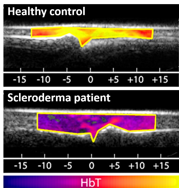
Reduction of perfusion in superficial tissue layers of a finger of a scleroderma patient, with total hemoglobin (HbT) shown as a pseudocolor overlay on a grayscale ultrasound image. Scales indicates position in millimeters.
Detection of microvascular dysfunction in systemic sclerosis
Systemic sclerosis manifests as fibrosis of the skin and internal organs and is associated with progressive microvascular dysfunction.
Optoacoustic imaging with MSOT provides the opportunity to study and quantify such dysfunction directly and non-invasively, as was done in a recent clinical trial.