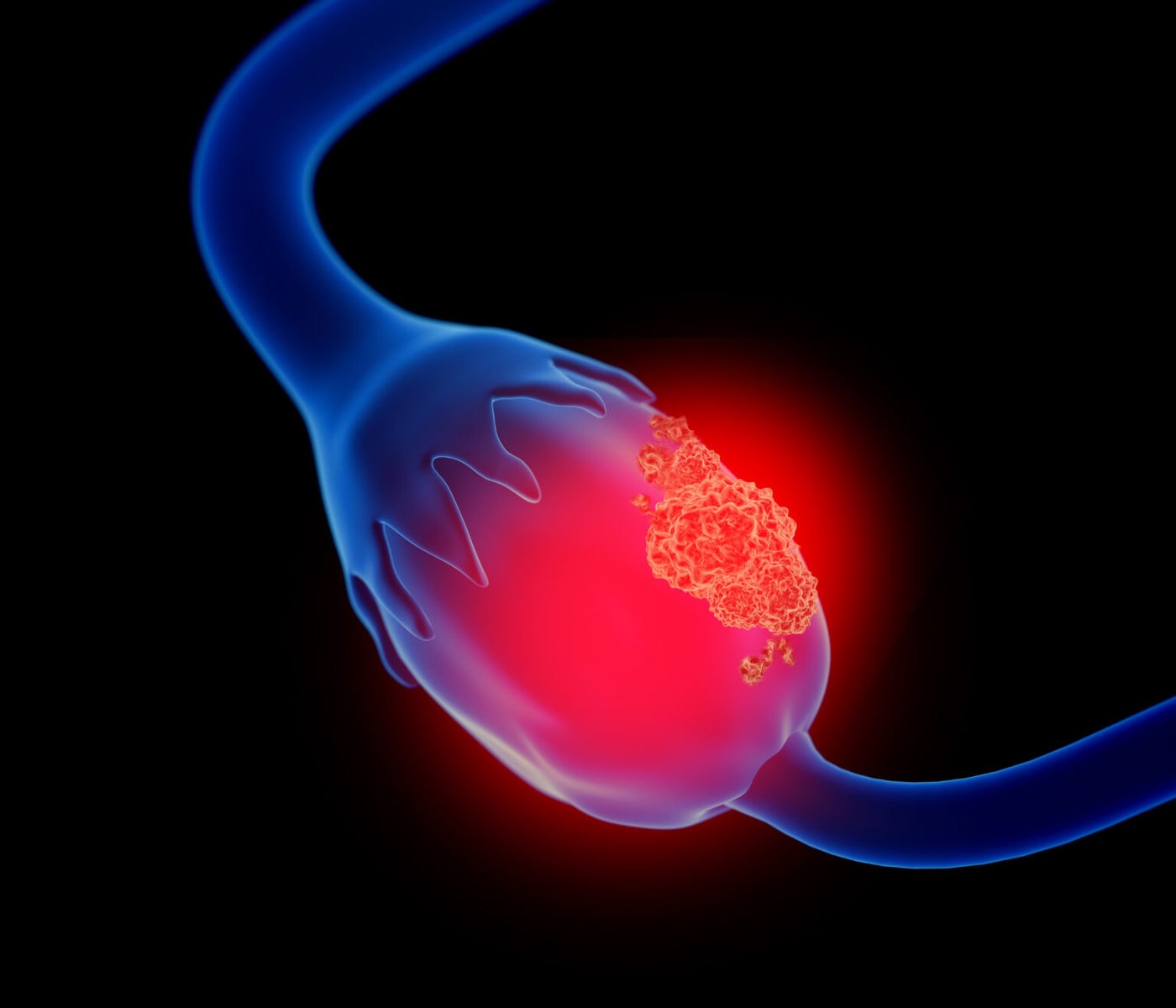
Breast cancer represents a significant burden to society. A particular risk is associated with diagnostic accuracy in patients with dense breast tissue. Initial studies have shown that optoacoustic imaging can potentially improve the specificity of diagnostic lesion work-up. Lesions are characterized according to changes in vascular morphology (angiogenesis) as well as changes in tissue composition (water/lipid ratio and distribution) and oxygenation (hypoxia).
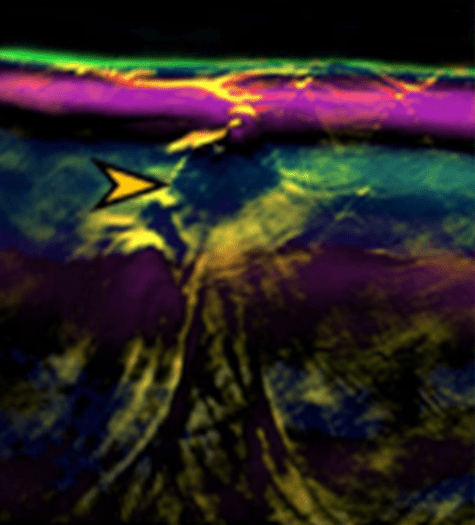
MSOT imaging of invasive lobular carcinoma (yellow arrow), visualizing the tissue chromophores water (blue), lipid (purple), and hemoglobin (with increasing oxygenation shown from green to yellow to red).
Novel assessment of malignant breast lesions
Recent clinical studies suggest that optoacoustic imaging could potentially fill this gap by providing additional anatomical and functional tissue information to distinguish benign from malignant breast lesions – thereby retaining the high sensitivity of ultrasound, while at the same time increasing the specificity.
Benign tissue generally shows homogenous optoacoustic signal associated with a homogenous breast tissue chromophore composition, particularly of hemoglobin, fat, and water. Conversely, cancerous tissue has distinct high-intensity patterns representing changes in breast tissue composition.
MSOT imaging is able to detect these differences in chromophore distribution and may provide additional anatomical and functional tissue information to distinguish benign from malignant breast lesions.
Diot et al. Clin Cancer Res. 2017
Observing tissue malignancy through vascular patterns
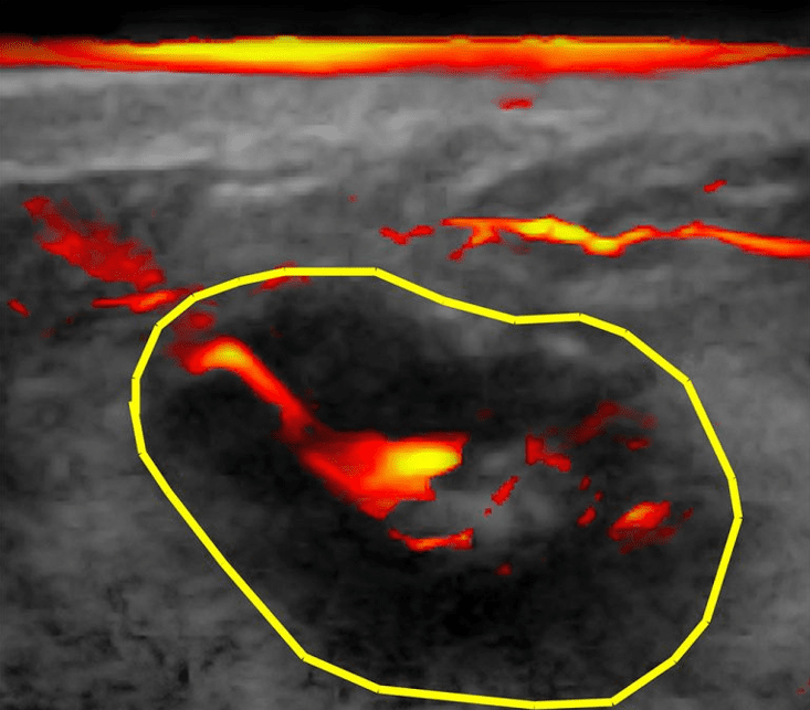
Besides changes in tissue composition, vascular patterns are often very indicative of malignant versus benign lesions; tumors tend to grow a vascular network that looks distinctly different from that in benign lesions, as large tumor-feeding vessels are frequently expressed in malignant lesions.
While initial results of investigator-initiated trials by MSOT users suggest that MSOT can help in improving the sensitivity and/or specificity of breast cancer imaging, further trials are needed to confirm this initial hypothesis. iThera Medical welcomes the opportunity to work with clinical users towards this end.
Adapted from MacCuaig et al. Radiol Imaging Cancer 2020
MSOT measurement of malignant breast lesion visualizing blood contrast from a tumor-feeding vessel overlaid on ultrasound background image