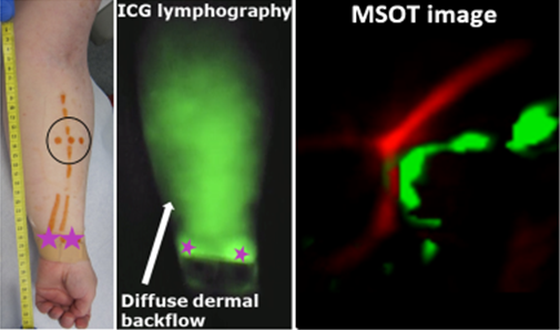
Visualization of lymph vessels is a necessary step in lymphatic reconstructive surgery. Indocyanine green (ICG) lymphography is currently the gold standard in preoperative lymphatic imaging; however, it can be limited by dermal backflow. MSOT combines the molecular specificity of optical imaging with the real-time aspects of ultrasound to provide high resolution imaging at depth. Endogenous contrast provided by hemoglobin enables bedside detection of the overlapping blood vascular network, and to select candidate sites for lympho-venous anastomosis.


ICG detection in lymphedema-affected limb via planar fluorescence and MSOT.
While lymphatic vessels filled with ICG and blood vessels can be distinguished based on the very different spectral profiles of ICG and hemoglobin, veins and arteries can be distinguished by spectral unmixing and by applying with the detector some pressure on the skin leading to the compression of veins but not of arteries.
Giacalone et al. J Clinical Medicine. 2020
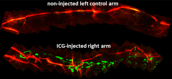
Images can be stitched together by moving the 3D detector along the length of the limb to provide a wider field of view spanning larger stretches of lymphatic and blood vessels. Lymphatic vessels had a segmented appearance over a length of 25cm along the forearm in agreement with ultra-high frequency ultrasound findings.
Giacalone et al. J Clinical Medicine. 2020

Maximum intensity projected images were stitched together thanks to overlapping areas in neighboring images.